
Research areas are arranged according to the laboratory's research framework. Publications for each area are listed in the same order on the Publications page.
A. Measurement and analysis
A1
Morphometry
Dimensions, areas, volumes and asymmetry are measured on three-dimensional surface models of regions such as the face, the whole trunk, the back and the extremities. Topics studied in this area include the effect of body position on facial soft tissue, the comparison of asymmetry calculation methods and the anthropometric accuracy of low-cost depth sensors. The methods are also applied to patient groups such as facial palsy, adolescent idiopathic scoliosis, Parkinson's disease, thalassaemia and stroke.
3D surface scanningAsymmetry (RMS)AnthropometryIntersection of morphometry and kinematics
Dynamic morphometry
Changes of the face during facial expressions are assessed by comparing three-dimensional scans taken at rest and during movement. The main topics in this area are the assessment of asymmetry and the monitoring of recovery in facial palsy, the quantification of facial movements in facioscapulohumeral dystrophy, the study of hypomimia in Parkinson's disease and the reliability of scanning methods.
Handheld 3D scannerFacial palsyFSHDA2a · Biomechanics
Kinematics
Facial and joint movements are measured by tracking passive reflective markers with an optoelectronic system. Topics studied include displacement and angular change during facial expressions, anatomical landmarks that remain relatively stable during movement, and shoulder range of motion. Depth sensors and markerless methods are compared against this system. Kinematic analysis of whisker movement in experimental facial nerve repair models in laboratory animals has also been carried out in this area using markerless systems.
This area also includes a wearable measurement system developed in the laboratory, which aims to track facial movement with flexible (flex) sensors placed on the forehead and cheeks. The study validating the sensors against synchronous optoelectronic recordings has been completed as an undergraduate student project; a manuscript is in preparation.
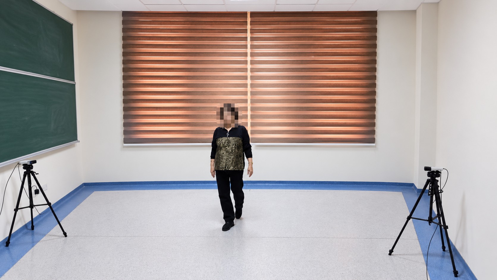
At the whole-body level, markerless gait analysis is performed with two Azure Kinect depth sensors and iPi Soft software. Joint angles are obtained from the recordings with the iPi Biomech add-on; a study of gait and lower-extremity tasks involving hip and knee movements in Parkinson's disease and in healthy individuals is ongoing.
A2b · Biomechanics
Kinetics
This area covers two levels of measurement: measurement of force and passive mechanical properties in living facial tissue with a dynamometer and, in the laboratory, study of the behaviour of biological tissues and materials under tension, compression and bending with a universal testing machine. Studies on the force and stiffness properties of facial tissue are ongoing.
DynamometerLloyd LR5K PlusTissue mechanicsA3
Neuromuscular activity
The electrical activity of facial muscles is studied with surface EMG recorded synchronously with motion analysis. Studies in this area include the relationship between surface morphometry, kinematics and muscle activity during smiling, and facial kinematics and muscle activity in different age groups.
Surface EMGSynchronous recordingB. Digital anatomy and model production
B
Digitisation and physical model production
Anatomical structures are converted into digital models with a 3D surface scanner or, from CT/MR images, with 3D Slicer. Physical models are produced from these by 3D printing, silicone moulding and liquid plastic casting.
Examples produced:
- Bone teaching models produced by silicone moulding and liquid plastic casting
- A pelvis model 3D-printed from a digital model created from MR images, intended to support preoperative preparation and anatomical orientation during surgery
- A patient-specific 3D-printed mould for shaping the PMMA implant in cranioplasty
- Sensor holders and calibration parts used in research
Son güncelleme : 2.11.2026 16:22:18